Cone Beam technology
Cone Beam technology
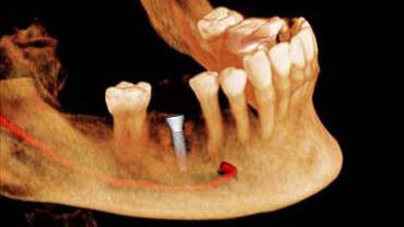
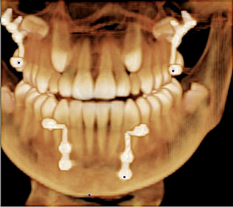
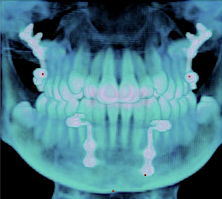
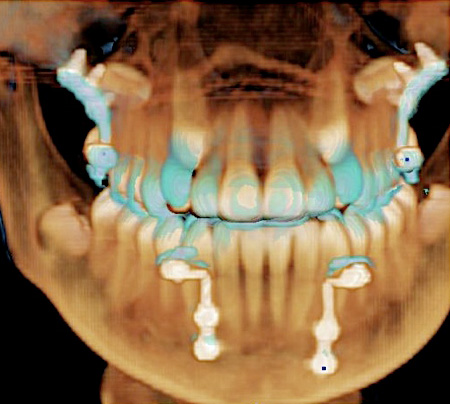
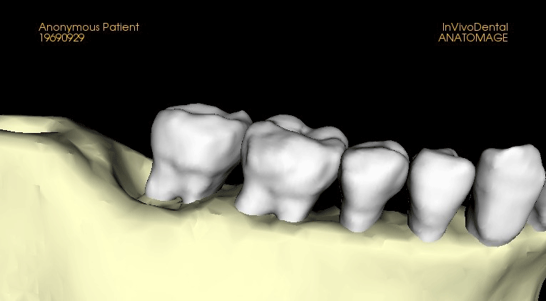
The introduction of Cone-Beam CT (CBCT) imaging for the maxillo-facial region has opened up new vistas for the use of three-dimensional (3D) imaging as a diagnostic and treatment planning tool for orthodontists, periodontists, implantologists, ENT physicians, and oral/maxillo-facial surgeons.
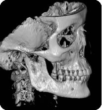
CBCT Scan using the NewTom VG Mobile Imaging Technology
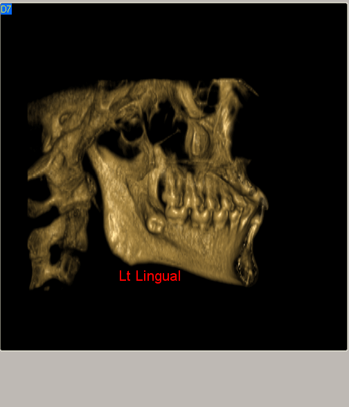
The three-dimensional imaging that CBCT generates allows for far more accurate diagnosis, treatment planning and monitoring, and analysis of outcomes than was possible with conventional two-dimensional images. Thanks to this revolutionary new imaging technology, coupled with our rapidly increasing understanding of the biological processes of maxillo-facial growth and development, we can now create and interact with virtual models of our patients’ tooth and jaw structures that enable us to provide a much higher level of treatment than ever before.
Data can be saved on CD, prints or access via Internet, and acts as an excellent tool for accurate diagnosis and more predictable treatment planning, reduces risks, enhancement of customer service and office prestige, more efficient patient management and education, improve treatment outcome, patient satisfaction and increase case acceptance.
Taking the lead in the development of this technology is Quantitative Radiology (QR), the company that invented dento-maxillo-facial CBCT technology. QR, a wholly owned subsidiary of AFP Imaging and the undisputed industry leader in dental 3D imaging technology, manufactures the NewTom family of CBCT imaging products. QR has consistently provided the highest quality imaging systems to the dental community through pioneering technological updates to its products and its continuing development of new products which advance the technology it invented.
CBCT vs. CT
Traditional CT (CAT Scan) uses a very narrow fan beam that rotates around the patient acquiring one or more thin axial slice(s) with each revolution. To image a section of anatomy, many rotations must be completed – which means high radiation exposure.
With Cone Beam 3D Imaging, rather than a narrow fan beam, a cone shaped beam is used to acquire the entire volume in a single pass around the patient, resulting in much less radiation exposure.
What is CBCT?
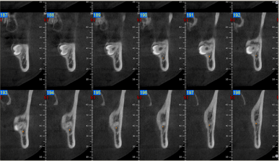
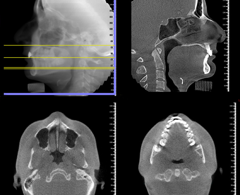
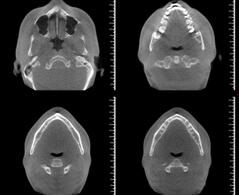
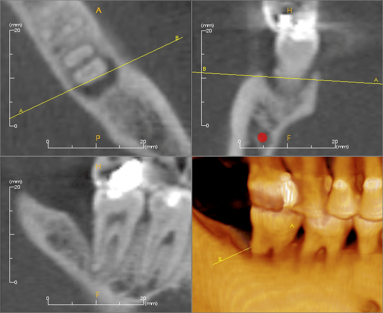
Cone Beam Computed Tomography is a technology specifically utilized for imaging the head, neck and jaws. Emitting cone-shaped x-rays, the scanner rotates around the patient’s head to capture a volume of high-resolution images. Digital files can be imported into 3-D viewing software such that the volume can be manipulated and examined in-depth from any view or angle desired. Axial, coronal and sagittal views can be viewed in life size so that accurate measurements can be taken. By adjusting the slice thickness of the images, critical anatomy such as nerve canals can be brought into focus.
Applications of CBCT include implant planning, oral surgery, orthodontic planning, TMJ, sinus and airway studies, and pathology.
CBCT imaging data can be saved on CD, prints, or accessed via the internet.
3D Volumetric vs 2D Imaging

3D Volumetric vs 2D Imaging
With 2D imaging, the letters are superimposed making it difficult to make out detail. With Volumetric Imaging, however, it is like removing a particular pane (slice) to examine it clearly and accurately.
Safe Beam™
Because the x-ray radiation is not on continuously, the 10-18 second duration of each scan translates to only 3.6-5.4 s of actually radiation exposure. A total of 360 individual images are taken, one per each degree of rotation.
Additionally, all NewTom systems incorporate Safe Beam™ technology, which automatically senses the size of the patient and drops the radiation dose by as much as 40% for younger, smaller patients, further reducing the total amount of radiation received.
Image reconstruction and reporting
When the scan has finished, the NewTom NNT software reconstructs the 360 images, transforming them into a three-dimensional database, representing the complete anatomy of the patient. This reconstructed volume consists of a series of axial images, the thickness of which can be graduated from 0.1 to 5 mm.
An unlimited number of these high-quality images can be constructed from the original volume, providing the operator with the ability to report on virtually any facet of the patient’s anatomy contained within the database. The NewTom software also allows you to measure, demarcate and visualize any aspect. You can distribute the reports using paper, videotape, email, CD or any other digital format. NewTom images can be acquired directly by Dolphin Imaging software, or outputted as a DICOM image for use with a wide variety of independent programs.
Imaging applications
Imaging applications
| Implantology Orthodontics Impactions |
Sinus and airway studies Pathology Endodontics |
TMJ Periodontics Oral surgery |
Implantology
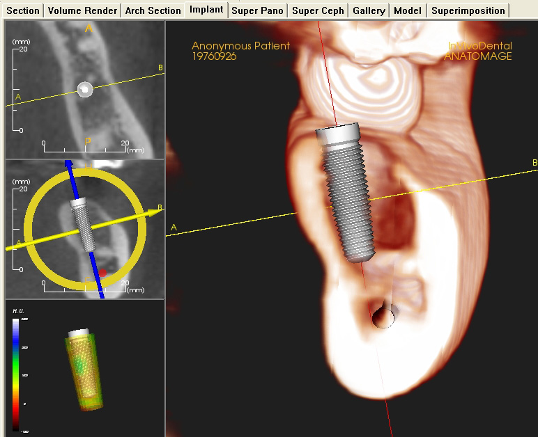
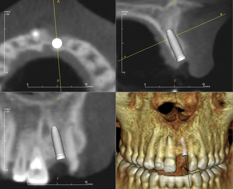
CBCT scans allow the surgeon and restorative dentist to optimally plan and place dental implants. Their uses and benefits are present throughout the continuum of care from diagnosis to treatment to post-op examinations and include:
- Locate and determine the distance to vital anatomic structures
- Measure alveolar bone width and visualize bone contours
- Determine if a bone graft or sinus lift is needed
- Select the most suitable implant size and type
- Optimize the implant location and angulation
- Increased case acceptance
- Reduced surgery time
- Build patient confidence
And with the use of guided implant placement based on CBCT scans, all the above benefits are enhanced to the point that the surgeon can approach each case with the confidence that comes from knowing that the best available image data and technology have been used to ensure success.
Orthodontics
Orthodontics has traditionally relied on 2-dimensional x-rays in evaluating 3-dimensional structures. With Cone Beam CT imaging, a more comprehensive orthodontic diagnosis and more accurate treatment planning is possible by allowing for:
- 3D views of vital structures
- 3D evaluation of impacted tooth position and anatomy
- TMJ assessments of condylar anatomy in three dimensions
- Orthognathic surgery treatment planning and growth assessments in true 1:1 imaging
- Airway assessments
- Planning for placement of dental implants for tooth restoration or orthodontic anchorage and for placement of temporary anchorage devices (TADs)
- Assessment skeletal symmetry or asymmetry
Impactions
Cone Beam CT scans can provide a more accurate and 3-dimensional assessment to provide more predictable treatment results while reducing the risks associated with any impacted tooth.
- Visualize an impacted tooth’s position in relation to surrounding vital structures and nearby teeth and their roots.
- Better assess the risk of treatment or non-treatment based on more accurate 3-dimensional analysis.
Sinus and airway studies
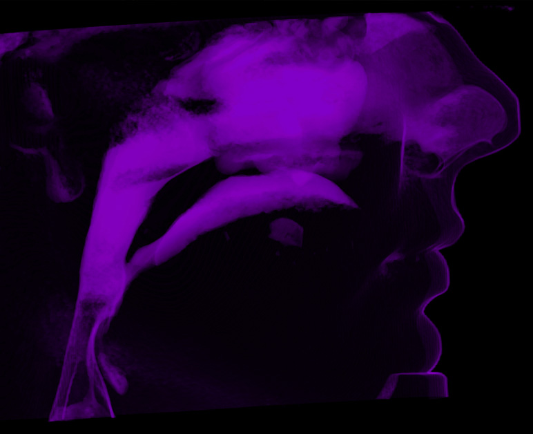
Volumetric data obtained from a CBCT survey can be used to visualize the sinuses and the entire airway path from the nasal and oral entrances to the laryngeal spaces for:
- Identification of anatomical borders
- Determination of degree of infection and presence of polyps
- Assistance in airway studies for diagnosis and treatment of obstructive sleep apnea
- Calculation of actual volume of airway space
- Determination of the point of airway constriction
Pathology
CBCT scans provide a superior means of visualizing and studying pathological processes in the maxilla and mandible. This information is invaluable when planning any surgical efforts for biopsy or resection. The data can be used to:
- Render three-dimensional images of hard tissue abnormalities
- Provide more accurate information related to size, extent, location, and the relation to and effect on nearby anatomical structures.
- Monitor the progression of the pathology as well as the success of treatment with the use of multiple scans.
Endodontics
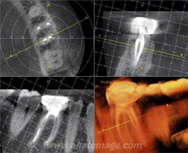
Although conventional radiography is more practical and better suited for everyday endodontic procedures, volumetric data from CBCT scans can provide serial axial, coronal, and sagittal views that are not possible to obtain from conventional radiography. The ability to reduce or eliminated superimposition of surrounding structures makes it easier to visualze areas of interest in three-dimensions. This provides much clinically relevant diagnositic information and has many potential applications for endodontics including:
- More accurate identification and diagnosis of periapical endodontic pathosis than conventional radiography
- Visualization of obscure internal pulpal anatomy and root canal systems
- Assessment of internal and external root resorptive processes
- Identification of root fractures and other areas of trauma
- Volumetric and density comparisons of periradicular bone following endodontic treatment in order to assess the degree of success or failure
- Pre-surgical planning
TMJ
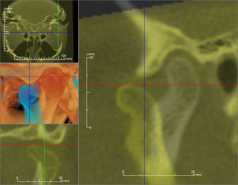
Accurate evaluation of the temporomandibular joint (TMJ) has been difficult due to the superimposition of other structures in conventional radiographs. With Cone Beam CT imaging, it is now possible to:
- Assess the condylar anatomy of the TMJ without superimposition and distortion of the image
- Obtain a true 1:1 imaging of the condylar structures for more accurate assessments.
Periodontics
The disadvantages of conventional 2-dimensional x-rays for accurate periodontal assessment is avoided by 3-dimensional and cross-sectional analyses helping to avoid surprises often encountered during periodontal surgery.
- Analyze periodontal bone defects on all sides of every tooth.
- Assess the extent of every furcation involvement.
- Track the progression of advancing periodontal bone loss.
- Treatment plan dental implants by evaluating bone parameters such as bone width, depth, and density.
- Visualize vital structures such as the maxillary sinus, mental foramen and mandibular nerve prior to periodontal or implant surgeries.
Oral surgery
In addition to implant placement, a Cone Beam CT scan is an invaluable diagnostic and treatment planning tool for the oral surgeon for:
- Determine the precise three-dimensional position of a tooth within the alveolar bone and how this position relates to vital structures for extractions and impactions.
- Visualize hard and soft tissues on the computer in three dimensions for planning maxillofacial surgeries.
- Monitor skeletal changes, airway changes, and healing responses.
Services
Services
NewTom CBCT
Since bringing Cone Beam CT technology to the dental arena in 2000, NewTom has continued to lead the industry in combining low dose patient exposure with high diagnostic, quality 3D images. NewTom’s foundation is built on a series of proprietary technologies which continue to leave NewTom products a step ahead of competition and in more professional imaging centers around the world.
All NewTom units utilize the patented Safe Beam™ technology, whereby low dose exposures capture scout scan data, and the tube head is automatically customized with the patient-specific adjustments. This eliminates any chance of operator error and adheres to the ALARA principal (as low as reasonably achievable). Safe Beam™ technology is responsible for up to a 40% radiation reduction to a child and frequently found in medical-grade imaging devices.
Flex Mobile Imaging Center version, feature variable FOV’s (15cmx15cm, 15cmx12cm, 12cmx8cm, 8cmx8cm and 6cmx6cm) at various different voxel sizes from .3mm down to .075mm. With various FOV and multiple voxel sizes to choose from, operators have the most options for image size and resolution specific to their specially – while always minimizing the dose accordingly.
A medical-grade tube head and small focal spot are responsible for the crystal-clear images for which NewTom is known. This outstanding image quality, resolution, and flexibility of variable FOV’s position the NewTom VGias the professional-grade choice for imaging centers and professional dental offices worldwide. Endodontists would utilize a smaller FOV than an Orthodontist or Oral surgeon would, so NewTom has built a solution for everyone into a single machine.
Focal Spot is a key factor in determining image resolution and quality of the X-ray image. As the focal spot decreases in size, resolution and the ability to detect detail improve. Because of its unique Rotating Anode, NewTom VGihas one of the smallest focal spots available. For even more precise measurements and treatment planning, the HiRes Zoom feature produces 50% higher image resolution and a 50% reduced slice thickness. Patient positioning features include a comfortable motorized chin rest and cross-hair positioning lasers.
An alternative way to utilize this “standard of care technology” is through the NewTom VGiFlex fleet of Mobile Imaging Centers. As the only machine which carries a warranty in a mobile environment, whether in your office or in a van, the NewTom VGiis built to stand up to the demands of professional diagnostic imaging anywhere the scan is taken.
VGi images are enhanced with greater contrast and less noise. Artifacts and scatter are negligible, leaving images, including soft tissue views, remarkably clean. NewTom VGitakes quality imaging to the next level
I-CAT Flex CBCT
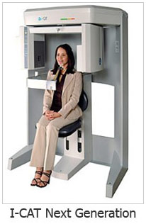 As the newest full field of view scanner developed by Imaging Sciences International, The I-CAT Flex is considered the state-of-the-art digital 3D scanner. It produces the highest quality images with a lower radiation dose than its predecessor, the I-CAT Flex, as well as other full field of view cone beam scanners on the market today. An additional advantage of the I-CAT Flex is the ability to reduce the field of view to as low as four centimeters.
The I-CAT flex’s high-definition, advanced imaging technology can be used by a variety of specialists, including orthodontists, periodontists, oral surgeons, implantologists, TMJ specialists, sleep specialists and general dentists.
The I-CAT Flex features an open environment seated position, a typical scan time of only 8.9 seconds and significantly lower radiation doses compared to traditional CT scans as well as other cone beam scanners on the market today. Since the scan time is reduced there is less potential for patient movement that can adversely affect the quality of the image produced. In addition, the immediate three-dimensional reconstruction of a patient’s mouth, face, and jaw areas enhances doctor/patient communication by allowing doctors to share a visual diagnosis with their patients. This gives patients a better understanding of their treatment options and more confidence when going into treatment.
The 3-D reconstruction gives doctors the opportunity to more thoroughly analyze bone structure, tooth orientation, and joint space, and detect and evaluate deformities and pathologies.
Cadent digital impressions
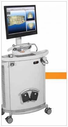
The iOC Cadent scanner, powered by iTero, is used to take full mouth orthodontic 3-D digital dental impressions. The Cadent iOC scanner eliminates the need to take dental impressions with impression material. These highly accurate, powder free, digital impressions are taken using laser and optical scanning technology.
Dental digital impressions can be used as a beginning, progress, or final orthodontic study model record. The models can be viewed and manipulated in the free OrthoCAD viewing software.
The Cadent iOC scan replaces the need for PVS Invisalign impressions. This highly accurate scan is sent directly to Invsalign who assigns it to the referring doctor’s account. The referring doctor can then log into the account and fill out an Invisalign prescription for the patient.
OrthoCAD iQ is a customized per-case service provided by Cadent. The iOC dental digital impression is sent directly to Cadent for computer-guided orthodontic treatment planning and bracket placement. OrthoCad iQ uses straight wire technique to calculate optimal bracket positioning for the referring doctor’s prescription. OrthoCAD iQ gives the referring doctor the ability to see post-treatment results on-screen before treatment proceeds. Using a virtual diagnostic setup, the OrthoCAD system places brackets to achieve the prescribed end result. After the setup is approved by the doctor, the pre-set brackets are delivered to the referring doctor in indirect delivery trays for bonding in the patient’s mouth. No dental impressions are necessary.
Radiology report
At the direction of the referring doctor, we can send any 2-D or 3-D radiographic image to be interpreted by a maxillofacial radiologist.
SimPlant master conversion
RDIC is the only master site in Wisconsin allowing us to format your dicom scans to SimPlant.
We offer a 24 hour turnaround for the formatting of your dicom.
Panoramic projection
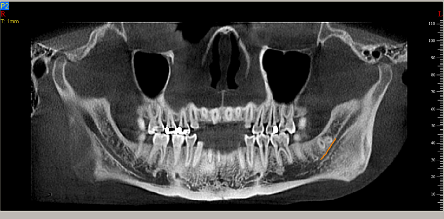
The panoramic is an overview image that is used to show the upper and lower jaws, erupted and unerupted teeth, and to evaluate root alignment, TMJ’s and the sinuses.
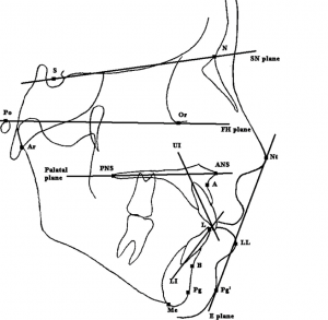
Cephalometric projection
These images are acquired in standardized lateral (side) and posteroanterior (front) projections. These views show the bone and soft tissues of the head, and are used to evaluate jaw growth and spatial relationships of the upper and lower jaws.
Cephalometric analysis
RDIC offers Cephalometric analysis.
Duplication services
RDIC offers duplication of 2D images.
Doctor FAQ
Doctor FAQ
How do I refer a patient to RDIC for imaging?
Please call 414-476-9400 to schedule an appointment for our Wauwatosa imaging center or call 414-943-7342 to schedule an appointment for our Mobile Imaging Center. You can also fax your referral form to 414-774-2803.
How are the appointments scheduled?
Appointments can be scheduled by the dental office staff or directly by the patient. Appointments can be made by fax, or by phone.
How soon will I get the images?
Immediately, or within 48 hours depending on time of day and complexity of service requested.
Can you convert the images to the planning software I own?
Yes, we convert to all 3rd party software. Please indicate on the referral form your preferred software.
Who should provide the scanning appliance?
The scanning appliance needs to be provided by the referring practitioner prior to the scan. It is essential that the practitioner try-in and adjust the template before the scan to insure accuracy.
How will I get training on the NNT viewer or I-CAT Vision software you provide?
One of our trained professionals will meet with you and your office staff to train, explain and demonstrate all features of the software. There is no charge for the viewer software or for the training.
What is the cost of the i-CAT Vision or NNT viewer software, and do you provide support for the software?
We provide the both viewer softwares at no cost. Training, full support and educational programs for the software at no additional cost. The viewer software can be utilized only for CBCT scans provided by RDIC.
Can I get a certified oral maxillofacial radiologist’s report?
Yes, for an additional fee, we provide you with a radiologist’s report within 3-5 business days. We send all requested scans to an oral maxillofacial radiologist who will render this service for you. This is the best possible medical-legal protection for you and a valuable health service for your patient.
Can I place virtual implants with the i-CAT or NNT software?
No. It is necessary to use third party software to place virtual implants such as Simplant, Nobel Biocare, Anatomage, etc.
Does the i-CAT or NNT provide visual and printed materials suitable for patient education?
Yes, either you, your dental treatment coordinator, or designated office staff person, can use the NNT online or printed images to demonstrate areas of pathology, virtual implants with opposing occlusion, impacted teeth, etc., which results in higher patient comprehension and case acceptance.
What are the additional benefits of RDIC?
Immediately or within 48 hours of the CBCT scan, depending on the time of day and complexity of services requested, we deliver a CD of scan, and/or printed images. A radiology report is available for a fee of $75.00 by a board certified oral maxillofacial radiologist within 3-5 business days, greater patient retention, increased office efficiency, patient management, and prestige, ultimate in customer service, most accurate diagnosis and treatment planning, improved treatment outcome and patient satisfaction, improved risk management, and higher case acceptance.
Who pays for the CT scan?
Payment for the CBCT scan is required the day of the scan by the referring doctor or the patient.
Do you provide measurements?
Yes, the professional software gives you the ability to do all precise and accurate measurements on your own. In addition, we provide you with a CD of the scan, and/or prints of the region of interest, if requested.
How much radiation (micro-sieverts) does the patient receive per scan?
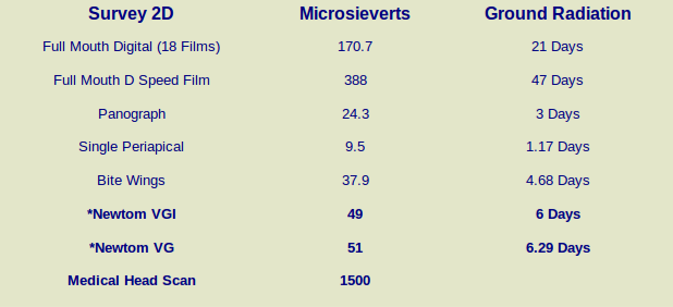
Cone Beam CT delivers less exposure than conventional studies but gives the referring doctor much more detailed information. Generally the patient will only have 1 or 2 scans during their visit depending on the image survey the doctor prescribes. In general, Cone Beam CT delivers only a radiation dose equivalent to two conventional panoramic x-rays. In comparison, a medical CT scan delivers 2200-3300 micro-sieverts, while a conventional panoramic x-ray delivers 20-100 micro-sieverts. The beauty of digital scanning, is that the huge volume of information is captured once and forever. From that volume the practitioner can reconstruct any view desired at any time without the need for further scans.
What am I able to see in Cone Beam CT scans?
Cone Beam CT allows practitioners to see anatomical structures with much detail. Anatomical anomalies such as the inferior alveolar nerve wrapped around an impacted third molar, bony dehiscences or fenestrations, questions about buccal/lingual structure locations, or even pathology are much more easily seen on a Cone Beam CT scan than conventional methods.
Referral form
Referral form
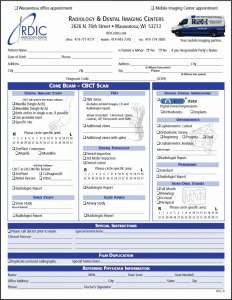 To order a scan for your patient, please download our PDF referral form,
then fill it out and FAX it to us at 414-774-2803.
When a x-ray assistant is utilized to perform a scan in the mobile unit, the ordering dentist assumes normal dentist responsibilities.
